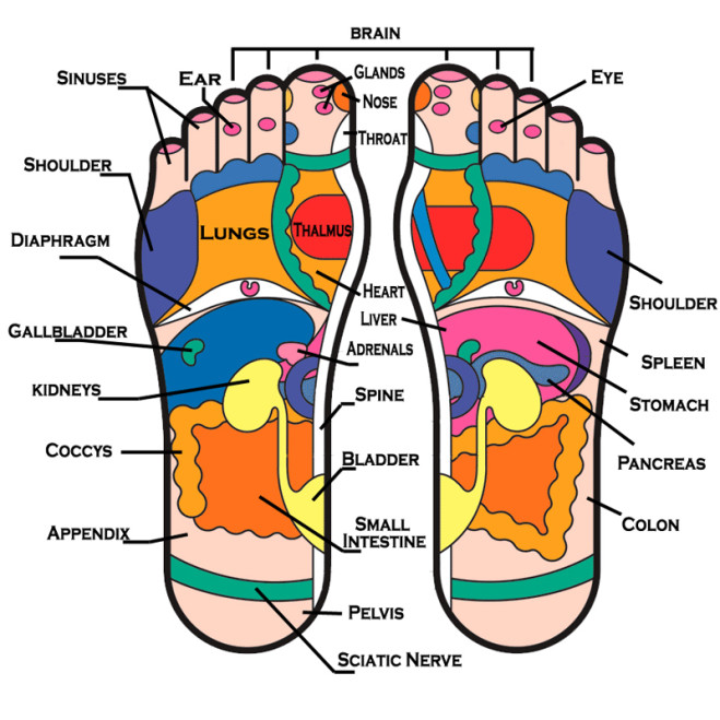
Sole Of Foot Diagram
Area soles toes human cutaneous were digits Fracture metatarsalgia joionline joi Diagram showing parts of the foot
Acute and Chronic Sciatica - Causes and removing the pain. | hubpages
Foot muscles sole layer third anatomy tendons bottom muscle netter medical netterimages ankle human labeled body pricing arthritis visit saved Underneath bottom underside plantar tendons ankle nerves fasciitis mikrora sponsored jooinn ligaments fascia Acute and chronic sciatica
What is metatarsalgia?
Nerves ankle tendon physiology tendons nerve ligaments organs leg skeleton sponsoredFoot muscles sole dorsal superficial lateral toes toe intrinsic left move right layer deep Foot chart reflexology parts feet body soles organs description internal corresponding previewSusan's blog: feet haven reflexology.
Chart organen lichaamsdelen beschrijving anatomie reflexology organs interne organReflexology foot chart charts feet massage map points sole pain oils acupuncture health bottom maps body therapy essential areas living Foot diagram sole under right vivian grisogono obliquely gardiner peter seenHurt wiring.

Foot sole area measurement. the surface areas of 9 different individual
Diagrams of footThis figure shows the muscles of the foot. the top panel shows the Diagram showing parts of the footFoot reflexology chart stock vector. illustration of physical.
The tibial nerveDiagram showing parts of the foot Muscles of sole of foot: third layerFoot sole innervation nerves sensory cutaneous nerve tibial toe anatomy lower which teachmeanatomy posterior diagram toes dermatomes leg motor innervate.

Diagram of your foot
Vivian grisogonoSciatica foot pain points sole causes acute nerve sciatic reflexology relief removing chronic Feet reflexology body parts chart areas foot sole different massage simple haven organs part bottom other health organ map linked.
.


